
(253) 236 – 5720

pacificretina@gmail.com

1412 SW 43rd St. Suite 206 Renton, WA 98057
(253) 236 – 5720
pacificretina@gmail.com
1412 SW 43rd St. Suite 206 Renton, WA 98057
Known as ARMD, Macular Degeneration can cause painless, loss of the central vision as we age. There are two main types of Macular Degeneration (Dry and Wet), and you may qualify for treatment depending on the nature/severity of your condition.
Direct trauma to the eye and/or orbit can cause damage to the sensitive tissues of the eye. If you have experienced significant trauma to your eye resulting in bruising, blurred vision, and/or pain, please seek medical attention immediately.
Diabetes can cause severe vision loss in several different ways. Leakage from damaged blood vessels can cause swelling in the central vision known as Diabetic Macular Edema. Damaged blood vessels can also impair blood flow (ischemia) to sensitive retinal cells. Bleeding in the eye (vitreous hemorrhage) can occur to due to the body’s attempt to repair damage by growth of new, harmful blood vessels (neovascularization).
Also known as cellophane maculopathy, epiretinal membrane can cause progressive, mild to moderate vision loss being associated with a history of trauma, ocular surgery, and inflammation of the eye. Most commonly, epiretinal membranes can arise spontaneously and can be observed without significant consequences.
A retinal detachment is the separation of the retinal tissue in the back of the eye away from it’s blood supply. There are several different types of retinal detachments including those caused by diabetes, systemic conditions, and trauma. There are also several methods to fix retinal detachments including in-office treatment (pneumatic retinopexy), scleral buckling (placing a band around the eye), vitrectomy (removal of the vitreous gel with use of a gas/oil bubble), and adjunctive laser treatment.
Pacific Retina Center
Mon - Fri 8:30 am - 5 pm
Sat - Sun *by appointment only
Phone: (253) 236 – 5720
Fax: (425) 988 – 0168
pacificretinacenter@gmail.com
1412 SW 43rd St. Suite 206 Renton, WA 98057
* Please allow 24 hrs for scheduling changes!
Q: What is the Retina?
A: The retina is the thin sheet of nerve tissue in the back of the eye containing light-sensitive rods and cones. Light that enters the eye is converted into electrical signals by the retina and these signals travel to the brain for processing to create sight.
Q: What will my first appointment be like?
A: Your first visit with us will involve a comprehensive eye evaluation, including dilated exam of your vitreous and retina. After carefully examining your retina, Dr. Ridgeley will educate you about your condition and treatment options, empowering you to make informed and confident decisions about your therapy.
Q: What kind of care will I receive?
A: Pacific Retina Center is a compassionate referral center dedicated exclusively to the diagnosis and treatment of retinal diseases. Dr. Ridgeley is committed to providing excellent care in a comfortable setting.
Q: Will my referral doctor or primary care physician be aware of my diagnosis/treatment plan?
A: Our team will work side by side with your doctor to ensure that you have the best possible care for your retinal condition.
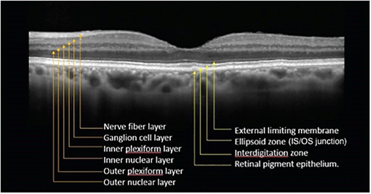
Optical coherence tomography (OCT) is a medical imaging technique that uses light to capture a high definition, 3-dimensional image of the optic nerve and retina. It is a non-invasive and non-contact technique way of taking a picture of structures within the retina to assist in the diagnosis of such eye diseases as macular degeneration, diabetic retinopathy, macular pucker, macular hole, optic nerve changes and central serous retinopathy.
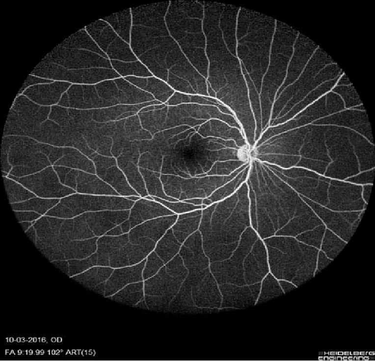
Fluorescein angiography is a specialized way of capturing high definition images using an injected dye to evaluate the structure and function of the blood vessels in the retina, choroid, optic nerve and iris. This simple, in-clinic test shows the possible swelling, leaking, and structural abnormalities resulting from such diseases as diabetic retinopathy, macular degeneration, blood vessel occlusions, infectious and inflammatory eye disease, tumors, and inherited eye diseases.
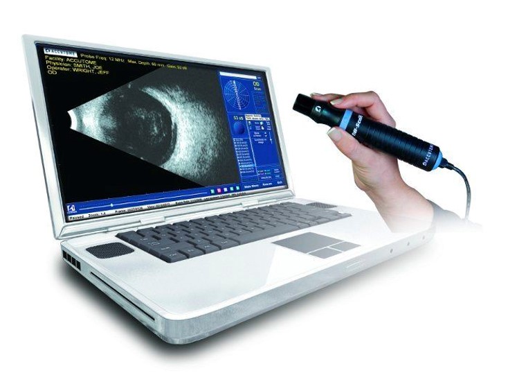
Ophthalmic ultrasound is a non-invasive method of imaging the front and back of the eye, as well as structures surrounding the eye. This type of ultrasound uses high frequency sound waves to capture 2-dimensional and cross sectional images of structures within the eye in situations of direct viewing through the eye is not possible, as in cases of bleeding in the eye, trauma and dense cataract formation.
Age Related Macular Degeneration
Diabetic Retinopathy
Retinal Detachments
Retinal Tears
Macular Edema
Macular Holes
Macula Puckers
Intraocular Tumors
Lucentis
Avastin
Anti-viral medications
Antibacterial medications
Eylea
Jetrea
© 2018 Pacific Retina Center PLLC. All Rights Reserved. Some 3rd party arts by vecteezy.com & Flaticon
By using this website, you agree to our privacy policies. and accessibility statement. Website developed by 1 Stop Link
Pacific Retina Center | Ophthalmologist in Renton, Seattle | Vitreo-Retinal Surgeon | Macular Degeneration | Ocular Trauma | Diabetic Eye Disease | Secondary Intraocular lenses | Retinal Detachment